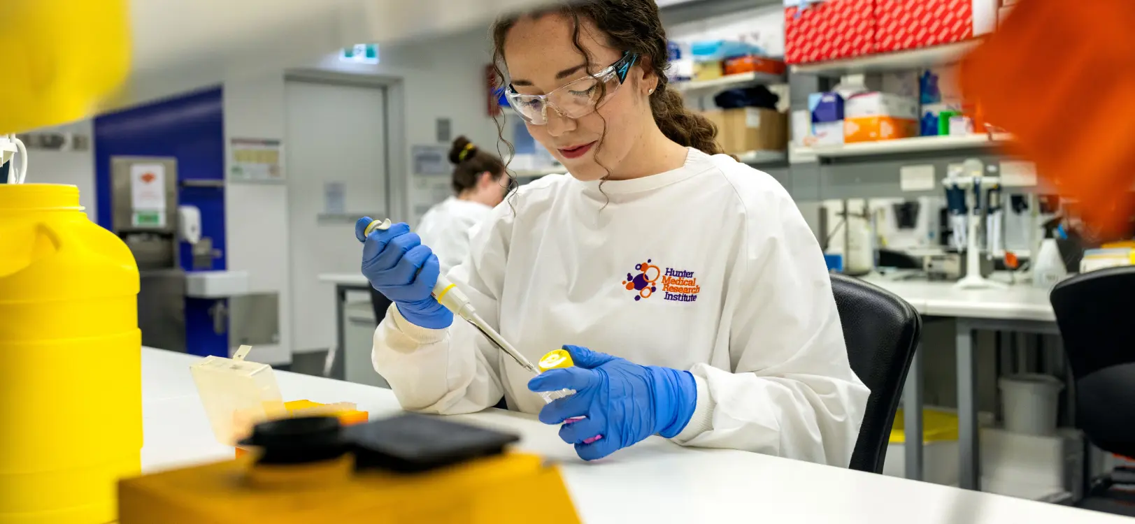
This behind-the-scenes video of the HMRI Histology and Biospecimen Resource Facility shows the processes involved in preparing a piece of tissue for biopsy from when it is collected, enters the laboratory, is processed, embedded, sectioned and stained for viewing underneath the microscope.
Have you ever wondered how slides for scientific research are created?
This behind-the-scenes video of the HMRI Histology and Biospecimen Resource Facility shows the processes involved in preparing a piece of tissue for biopsy from when it is collected, enters the laboratory, is processed, embedded, sectioned and stained for viewing underneath the microscope. This process is crucial in developing an understanding of the makeup and composition of pieces of tissue.
The HMRI Histology team processes a range of tissue samples for gynaecology research, respiratory, gut and various cancers including brain, breast, lung and colon.
Dr Melissa Tadros from the School of Behavioural Sciences and Pharmacy at the University of Newcastle created this video as a student resource, so they could understand the process and the steps involved in preparing a piece of tissue for biopsy.
- The piece of tissue is received in a small vial with 70% ethanol. It’s placed into a processor which removes the ethanol and embeds the tissue in paraffin wax. The processor has programs for different kinds of tissue.
- The wax containing the tissue is cut into sections 4 microns thick.
- These thin ribbons of the wax are placed in a hot water bath and then floated onto slides.
- Multiple stains or dyes are put on the tissue so it can be viewed under a microscope. The dyes give a good representation of the overall architecture of the tissue.
The lab has several hi-tech machines including a scanner that can process up to 400 slides per session. Slides can be viewed up to a 40x magnification level and the resulting images put online and shared with collaborators anywhere around the world.